|
||
|
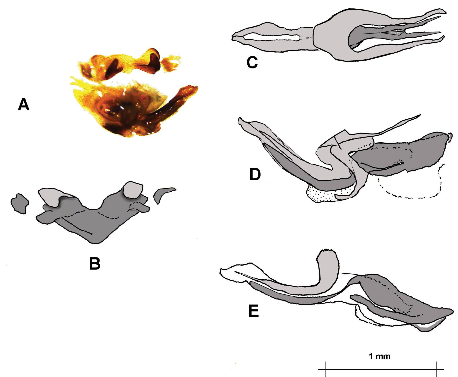
Mopla guttata, phallic structures. A. Oblique posterior view of phallic complex before preparation and dissection; B. Epiphallus, anterior view; C. Dorsal and D. Lateral views of phallic complex with epiphallus, epiphallic, and ectophallic membranes removed; and E. Endophallus, arch sclerite, and ectophallic aedeagal valves, after removal of remaining ectophallic structures. In C–E the endophallus is in a darker shading, the ectophallus in lighter shading. The broken line in D indicates the presumed position of the ejaculatory sac, missing from this preparation. Spermatophore sac stippled. |