|
||
|
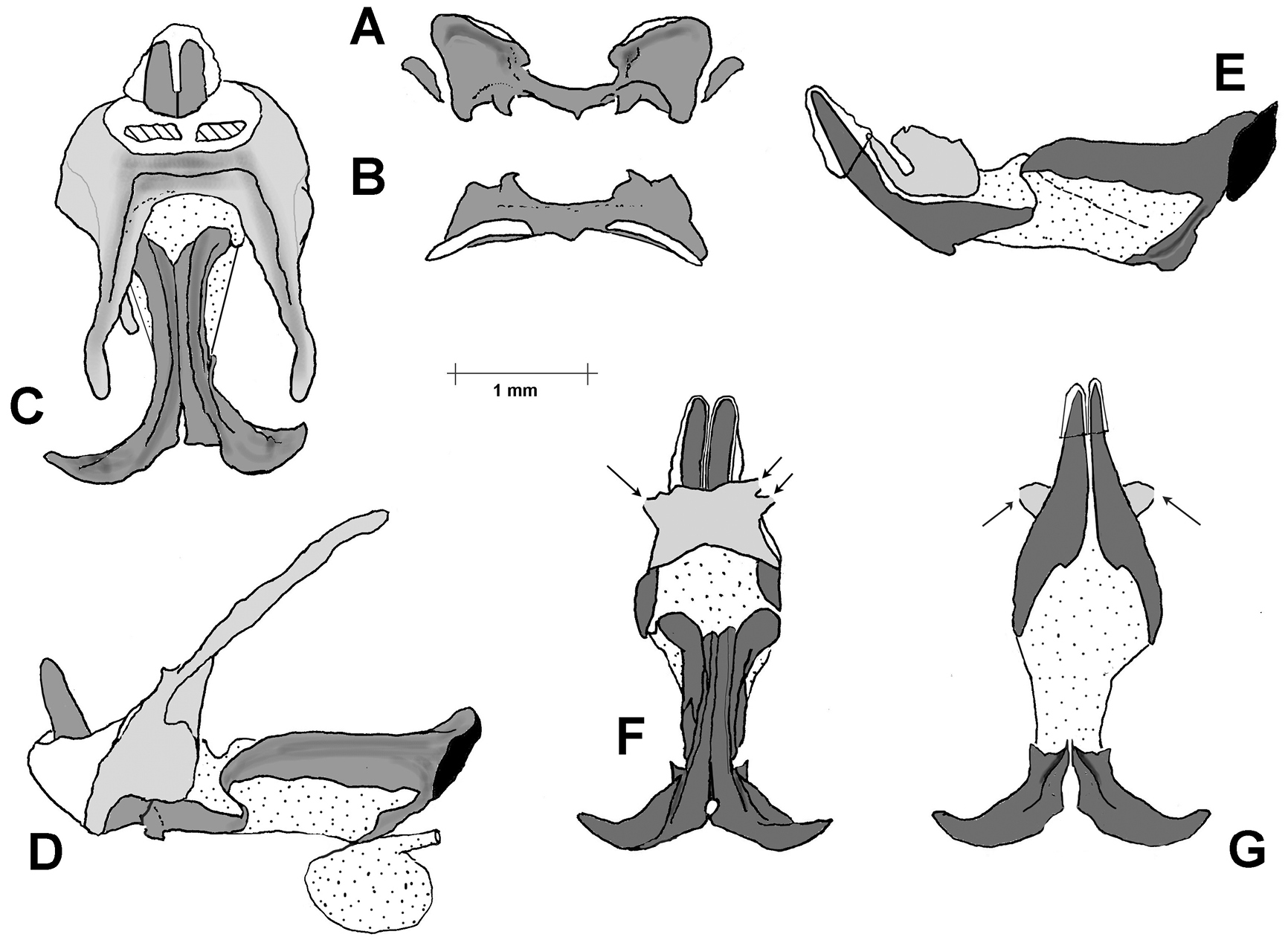
Serpusia succursor, phallus. A. Epiphallus, axial view; B. Epiphallus, dorsal view. C. Phallic complex after removal of epiphallus and epiphallic membrane, dorsal view. The two cross hatched areas are the zones of attachment of the the arch to the inner surface of the zygoma; D. As in C, but lateral view; E. Endophallus and arch in lateral view; F. As in E but dorsal view. The arrows indicate cut margins of the arch where it has been freed from the inner surface of the zygoma; G. As F, but ventral view. In E, F and G the ejaculatory sac has been removed to show the detail of the gonopore processes. |