|
||
|
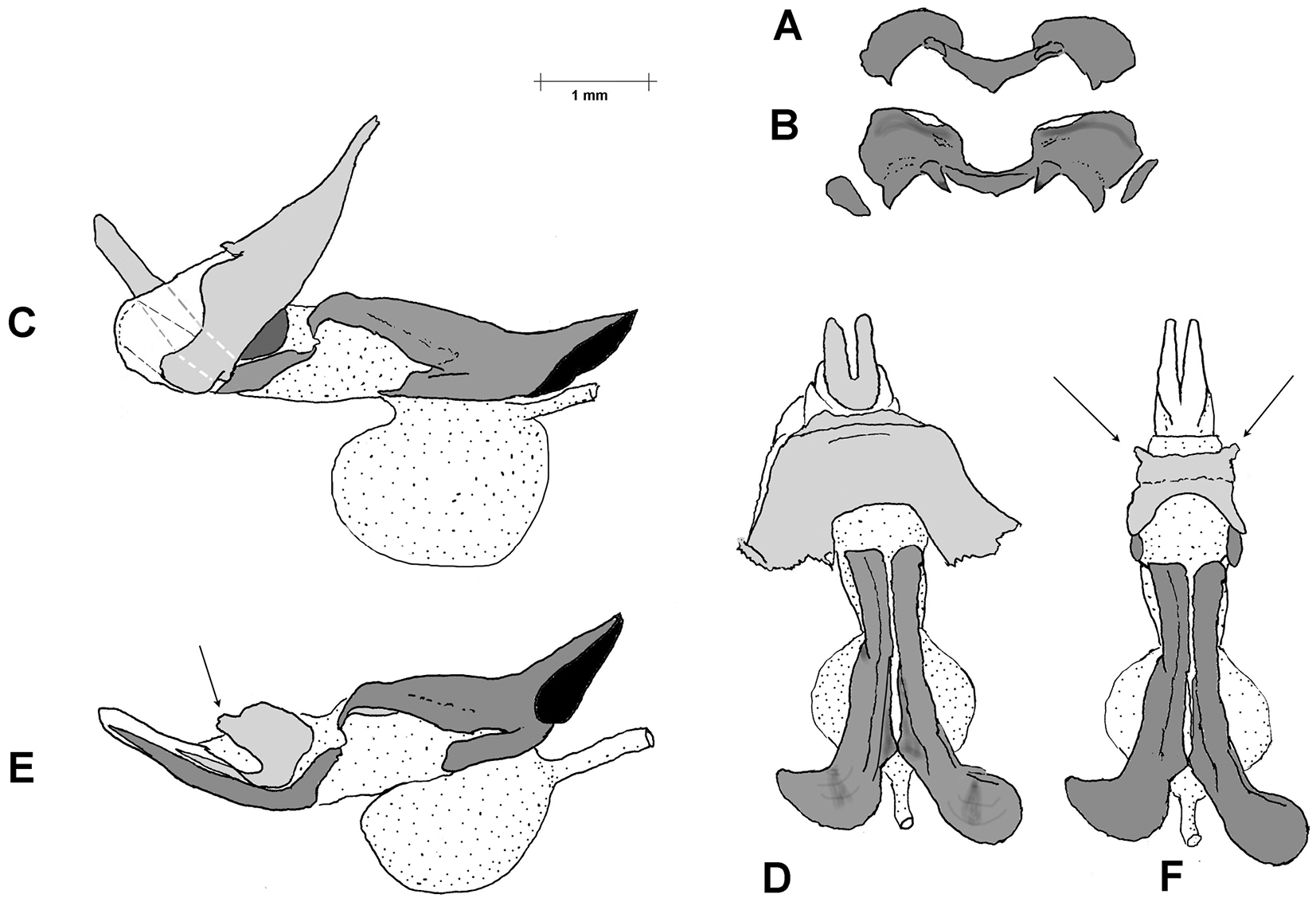
Serpusia opacula, phallus. A. Epiphallus, axial view; B. Epiphallus, dorsal view; C. Phallic complex after removal of epiphallus and epiphallic membrane, lateral view; D. As in C, but dorsal view. In this preparation the cingular apodemes are broken off; intact, they resemble those of S. succursor (Fig. 8), being long and straight and almost parallel; E. Endophallus and arch, lateral view; F. As in E, but dorsal view. In E and F, the arrows indicate cut margins of the arch where it has been freed from the inner surface of the zygoma. |